Figure 1.
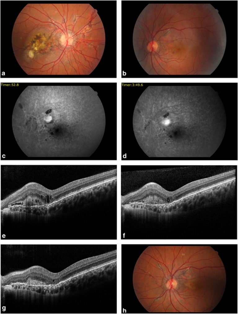
Fundus photograph befor aflibercept injection (a) AS with macular disciform skar in the right eye, (b) AS with multiple intraretinal hemorrhages at the extrafovea region in the left eye. (c, d) Fluorescein angiography proved extrafoveal classic CNV with an active leakage on the early and late phase in the left eye. (e) Optic coherence tomography showed intraretinal fluid before aflibercept treatment. (f) OCT revealed decreased intraretinal fluid 1 month after first injecton. (g) After loading dose there was resolution of the intraretinal fluid. (h) Fundus photograph after loading aflibercept dose showed resolution of the intraretinal hemorrhages.

