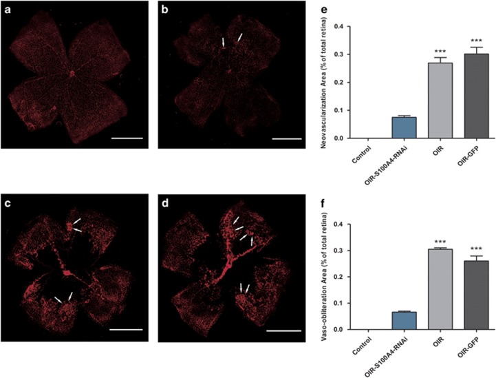
Figure 3.
Effects of Ad-S100A4-RNAi transfer on oxygen-induced ischaemic retinal neovascularization. Immunofluorescence staining of vasculature on retinal flat mounts at P17. (a) Control group; (b) OIR-S100A4-RNAi group; (c) OIR group; (d) OIR-GFP group; 40 × magnification. The white arrows indicate the neovascular tufts. (e) Quantification of the neovascularization area in the retinas from the OIR-S100A4-RNAi group, OIR group, and OIR-GFP group. Retinal neovascularization was quantified by measuring the ratio of the neovascular tuft area to the total retinal area using Image J 1.48V software (NIH; mean±SD). OIR-S100A4-RNAi group vs OIR group, ***P<0.05; OIR-S100A4-RNAi group vs OIR-GFP group, ***P<0.05. (f) Retinal vaso-obliteration areas were measured by calculating the ratio of the non-perfusion area to the total retinal area using Image J 1.48V software (NIH; mean±SD). OIR-S100A4-RNAi group vs OIR group, ***P<0.05; OIR-S100A4-RNAi group vs OIR-GFP group, ***P<0.05.

