Figure 3.
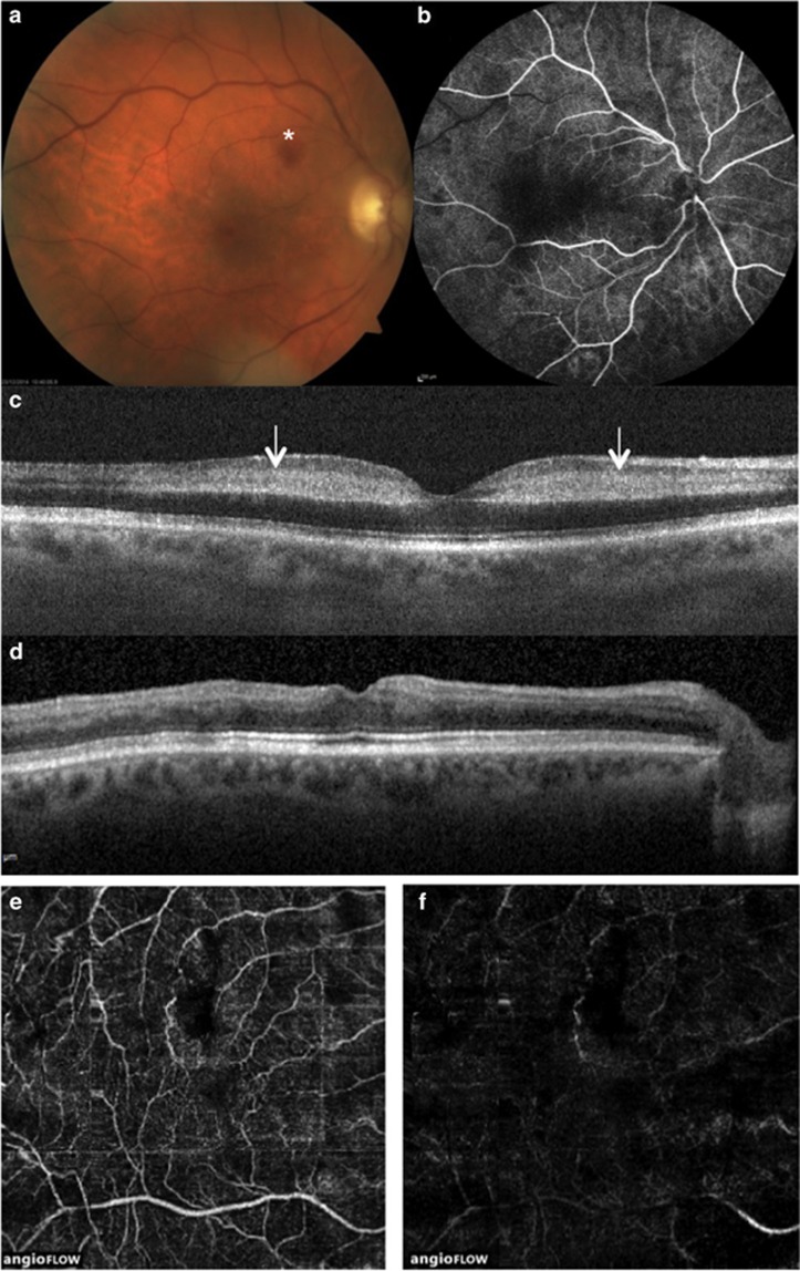
Case 3. Multimodal imaging of right eye. BCVA at presentation was 26 ETDRS letters; CFP (a) revealed venous engorgement and a retinal hemorrhage at the posterior pole (*); filling was slow during venous phases of FA (b), which revealed no areas of capillary closure; SD-OCT scan (c) showed hyperreflective band-like lesions involving the middle layers of the retina at the level of the INL (arrows). After 9 months, BCVA was eight ETDRS letters; SD-OCT scan (d) revealed marked thinning of the INL; OCTA showed mild attenuation of the SCP (e) and extensive capillary dropout of the DCP (f)

