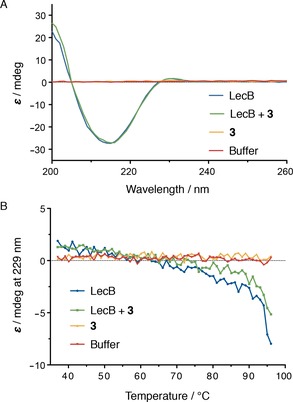
Figure 6.

A) CD spectroscopy of LecB (35 μm) in the presence or absence of compound 3 (100 μm) gave identical CD spectra indicative for beta‐sheet secondary structure. B) thermal unfolding followed by CD spectroscopy at 229 nm was not indicative of a destabilization of the protein in presence of 3.
