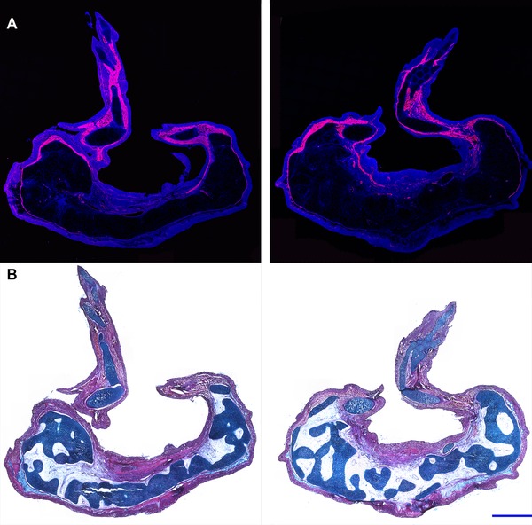
Figure 2.

Histology of RA‐induced paired limbs from an anterior wound site. Histological analyses were performed on sections that transected each of the two ectopic limbs that grew from an RA‐treated anterior wound site, harvested 10 weeks post‐treatment. Two complete limbs, including the zeugopod, stylopod, and autopod, formed from this experimental manipulation. A large mass of tissue formed proximal to the limb structures. (A) Fluorescent images were obtained of the cryosectioned limbs stained with DAPI (blue) and phalloidin‐rhodamine (red). Muscle tissue, rich in F‐actin (stained with phalloidin‐rhodamine), was distributed throughout the more distal limb structures but was not observed in the mass of tissue located proximally. (B) Sections were stained with eosin Y, hematoxylin, and alcian blue for histological analysis. The proximal mass of tissue predominantly differentiated into connective tissue and cartilaginous elements (dark blue) that could not be identified as corresponding to skeletal elements that are part of the normal limb pattern. Blue scale bars are 1 mm in length.
