Figure 3.

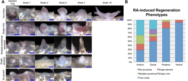
RA‐induced regeneration phenotypes are position‐specific. Quantification of the regeneration phenotypes for RA‐induced blastemas. Upon the completion of differentiation, the ectopic outgrowths were collected and stained for bone and cartilage in whole mount preparations in order to analyze the presence of skeletal elements in the regenerate. From our observations of these skeletal preparations, we divided the regeneration phenotypes into five categories: (1) no ectopic structures; (2) a single skeletal element; (3) multiple (jointed) symmetrical skeletal elements; (4) a single limb; or (5) two paired limbs (also see Table 1). (A) Examples of blastemas that exhibited the five different regeneration phenotypes that were quantified in this study. Images were taken of the blastemas starting the day of RA injection and ending when the tissue in the wound site had completely differentiated (determined by the formation of mature skin). (B) The histogram represents the percentage of ectopic blastemas located on the anterior, dorsal, posterior, or ventral axis that differentiated into each phenotype. Only the wounds located on the anterior or dorsal axis generated one or two paired limbs when treated with RA (also see Table 1). Blue scale bars are 1 mm in length.
