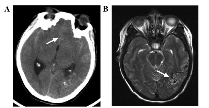
Figure 1.

CT and MRI imaging of the head of the patient. (A) CT scan showed a high-density cord-like shadow in the lateral Sylvian cisterns and the longitudinal Sylvian (white arrow), suggesting a subarachnoid hemorrhage. (B) MRI scan revealed a flow-void signal in the left occipital lobe (white arrow), suggesting arteriovenous malformation. CT, computed tomography; MRI, magnetic resonance imaging.
