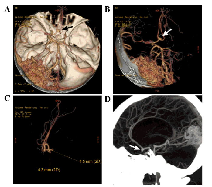
Figure 3.

Computed tomography angiography image of the head of the patient. (A and B) An AVM was evident in the left occipital lobe. The branches of the left middle cerebral artery and posterior cerebral artery can be observed entering the lesion. The draining veins from the lesion converged into the superior sagittal sinus upward and the transverse sinus backward. The right middle cerebral artery and the anterior cerebral artery A1 segment were not normally displayed, and the left anterior cerebral artery A2 segment started from the anterior communicating artery (black and white arrows). A blood vessel branch in close to the left middle cerebral artery extended into the AVM. (C and D) An aneurysm of ~4.6×4.2 mm was observed in the anterior communicating artery (white arrows), projecting to the lower right side. AVM, arteriovenous malformation.
