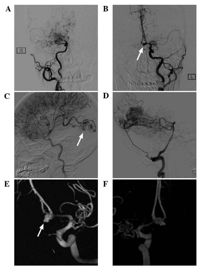
Figure 4.

Digital subtraction angiography image of the head of the patient. (A and B) Carotid angiography showed that the bilateral middle cerebral artery was not present and was replaced by abnormally developed moyamoya-like vessels, and the right anterior cerebral artery was absent. The left anterior cerebral artery was dominant, serving the bilateral anterior cerebral artery, and an aneurysm was present in the anterior communicating artery (white arrow). (C and D) An arteriovenous malformation in the occipital lobe was observed, with the blood supply from the middle cerebral artery and the posterior cerebral artery (white arrow) and the draining veins converging into the superior sagittal sinus upward and the left transverse sinus backward. (E and F) 3 dimensional reconstruction showed that the anterior communicating artery aneurysm had an irregular shape, with ruptured vesicles present on top (white arrow).
