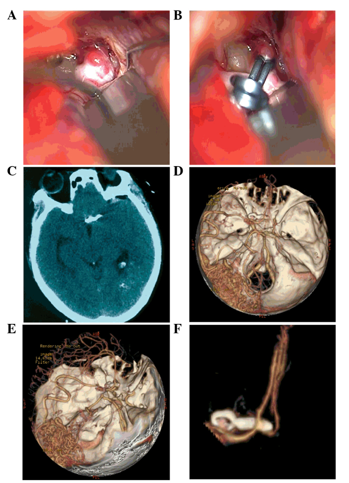
Figure 6.

Intraoperative and postoperative images of the head of the patient. (A) A preoperative image of the anterior communicating aneurysm. (B) A postoperative image of the clipped anterior communicating aneurysm. (C) A postoperative computed tomography scan showed intact morphology of the brain tissue. (D-F) The follow-up computed tomography angiography six months after the surgical procedure showed a good result for the aneurysm clipping, with no changes in Moyamoya disease or arteriovenous malformation.
