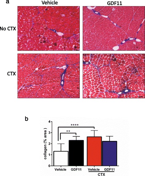
Fig. 4.

Assessment of fibrosis upon GDF11 treatment. a Representative images of the TA muscles stained with Masson’s trichrome. The presence of blue between myofibers represents collagen content, which is indicative of fibrosis. b Quantification of fibrosis shows GDF11-treated mice present increased levels of muscle collagen content in comparison with vehicle-treated mice. As expected, collagen content increased upon CTX injury, however, only in vehicle-treated mice. Scale bar = 100 μm. **p < 0.01, ****p < 0.0001
