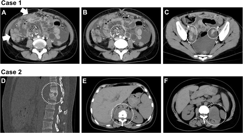
Fig. 2.

Abdominal CT findings of case 1 (a–c) and case 2 (d–f) at diagnosis. a, Moderate ascites and intraabdominal mass lesions (arrows). b, intraabdominal lymph nodes swelling (in the circle). c, bilateral adnexal masses (in the circle). d and e, Osteosclerotic lesion of Th11-Th12 vertebra with surrounding abscess (in the circle). f, A large low attenuation mass in the left psoas muscle (in the circle)
