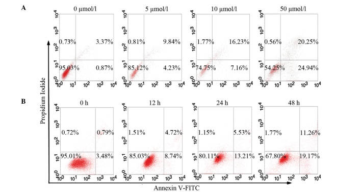
Figure 2.
CecropinXJ induced apoptosis in Huh-7 cells. Huh-7 cells were treated with (A) different concentrations of cecropinXJ (0, 5, 10 and 50 µmol/l) for 24 h, or with (B) 10 µmol/l cecropinXJ for 0, 12, 24 and 48 h. Cells were stained with Annexin V-fluorescein isothiocyanate and propidium iodide, and analyzed by fluorescence-activated cell sorting. The number of apoptotic cells (Annexin V+) was indicated as the percentage of gated cells. (B) The percentage of apoptotic cells was expressed as the mean ± standard deviation of triplicate samples. FITC fluorescein isothiocyanate, PI, propidium iodide.

