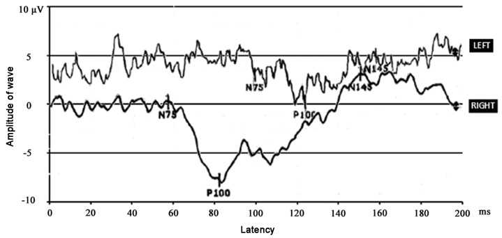
Figure 1.
Visual evoked potentials (VEPs) obtained at presentation upon left (upper waveform) or right (lower waveform) eye stimulation (1 Hz contrast reversal of 1 degree black and white checks). VEP obtained upon left eye stimulation was delayed and reduced in amplitude. Major components of the response (N75, P100 and N145) are indicated.

