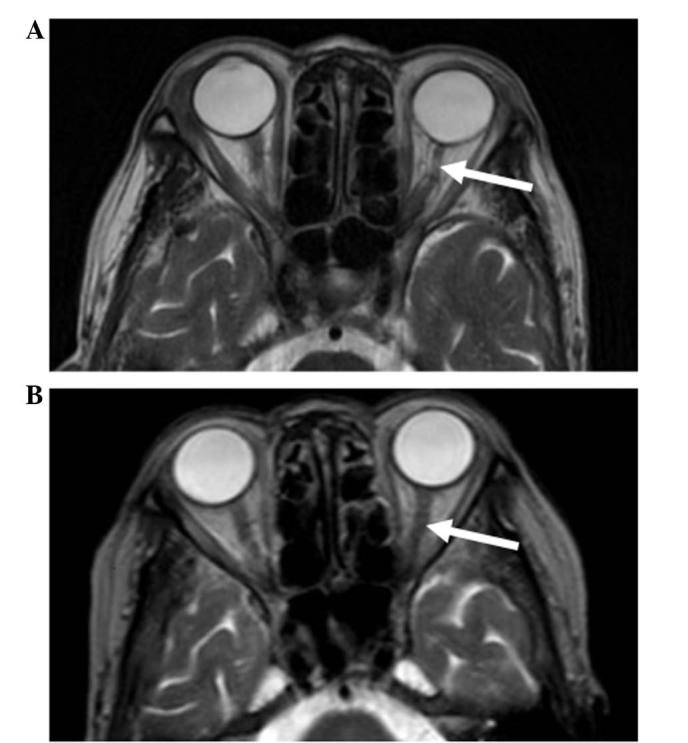
Figure 2.

Cranial magnetic resonance T2-weighted images obtained: (A) At presentation, showing a thinner left optic nerve (arrow) compared with the right optic nerve, and presence of a hyperintense lesion; and (B) after 4 months of oral methylprednisolone therapy, showing that the appearance of the left optic nerve (arrow) was similar to that of the right optic nerve and the hyperintense lesion had resolved.
