
Figure 3.
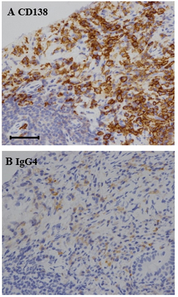
Histopathological findings in cervical lymph node biopsy. (A) CD138 immunohistochemical staining (brown) identified plasma cellular infiltration in the cervical lymph node (magnification, ×400). (B) Immunoglobulin G4 (IgG4) immunohistochemical staining (brown) labeled a large number of IgG4-positive plasma cells among the inflammatory infiltrates (magnification, ×400). The ratio of IgG4/IgG cells was >40%, which was much higher compared with the 6% ratio typically observed in normal subjects. Scale bar=50 µm.