
Fig. 4.
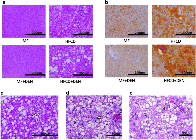
Representative images of stained liver sections: a 12 weeks with hematoxylin-eosin staining; b 12 weeks with Sudan staining; c–e 16 weeks in HFCD + DEN mice with hematoxylin-eosin staining. The original magnification is × 200 (a–c) and × 400 (d). Lipogranuloma (c), a Mallory-Denk body (d), and hepatocyte ballooning (e) are indicated by yellow arrowheads. MF, standard diet; HFCD, high-fat choline-deficient diet; DEN, diethylnitrosamine