
Fig. 6.
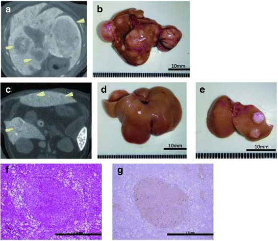
Computed tomography scans and immunohistochemistry of hepatic tumors in the high-fat, choline-deficient (HFCD) + diethylnitrosamine (DEN) group at 24 weeks: a, c computed tomography findings; b–e macroscopic views. The image in a is a section of the whole liver depicted in b; the image in c is a section of the whole liver shown in d; and the image in panel e depicts the right and left medial lobes of the whole liver in panel d. The lesions are indicated by yellow arrowheads. f Hematoxylin-eosin staining of the liver tumor. g Immunohistochemical staining for glutamine synthetase