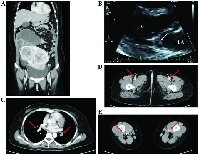
Figure 2.
(A) Contrast-enhanced computed tomography (CT) scan revealed a 20-cm multiple uterine myoma and a 10-cm right adnexal tumor that was suspected to be an ovarian malignancy. (B) Transthoracic echocardiography showed vegetations on the mitral valve (arrow). The largest area of vegetation, 7 mm in length, was attached to the anterior leaflet. LA, left atrium; LV, left ventricle. (C-E) CT scan showed (C) emboli in multiple pulmonary arteries (arrows) and (D and E) thrombus formation in the lower limbs bilaterally (arrows).

