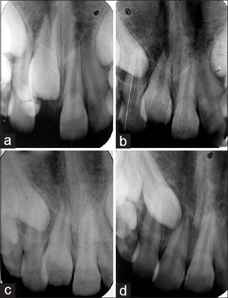
Figure 7.

Intraoral periapical radiographs of the erupting central incisor and change in path of eruption of erupting canine during follow-up of 2 years. (a) Preoperative intraoral periapical radiograph showing impacted central incisor and presence of two tooth-like denticles. (b) After 1 year follow-up showing movement of impacted maxillary central incisor. (c) Intraoral radiograph showing 1. years of follow-up. (d) Radiograph showing complete eruption of impacted incisor after 2 years
