Fig. 2.

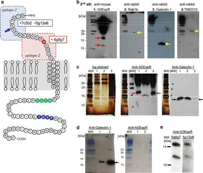
Analysis of DEspR translatability. a Schematic diagram of DEspR protein and mAb-epitopes. Two distinct peptides (epitope-1, epitope-2) in the extracellular domain were used to develop murine monoclonal antibodies (mAbs). Two high-affinity mAbs target human-specific epitope-1: 7c5b2, 5g12e8; and one high-affinity mAb targets the pan-species reactive epitope-2, identical in human, monkey, and rat. Epitope-2 spans the putative ligand binding domain [24]. The 5g12e8 mAb was used in pull-down experiments; 5g12e8 and 6g8g7 were used in Western blot analyses, 7c5b2 mAb was used in FACs analysis, immunostaining, and internalization assays, and all three were used in functional inhibition assays. The contested tryptophan (W)-aa#14 (red); consensus glycosylation site sequence: (green, N-F-S-G), known internalization recognition sequence: (blue: T-D-V-P). A blue arrow marks the splice junction between exon1 and exon2, i.e. between amino-acids G and K (aa#5-#6). b Sequential Western blot analyses of pull-down proteins from glioblastoma U87 membrane proteins using different antibodies specific for proteins identified by mass spectrometry analysis of pull-down protein-products. The identical blot was sequentially probed, stripped of antibody, confirmed as stripped, then re-probed in the following order: #1: anti-hDEspR-5g12e8 mouse mAb, #2: anti-Rab1b rabbit polyclonal Ab (pAb), #3: anti-Galectin-1 rabbit pAb; #4: anti-TMED10 rabbit pAb. Molecular weight markers are noted. DEspR bands are ~17.5 and 12.5 kDa. Expected sizes are detected for Rab1b: 22 kDa, Galectin-1: 14 kDa, and TMED10: 25 kDa. c Panel-1 shows silver-stained gel of pull-down protein products using 5g12e8 mAb from membrane proteins isolated from: (1) glioblastoma U87 CSCs, (2) PNGase-digested sample of pull-down proteins from U87 CSCs, (3) permanent transfectants DEspR-positive Cos1-cells. Panel-2 shows Western blot analysis using anti-DEspR 5g12e8 mAb showing DEspR band (lane 1), smaller DEspR + band after PNGase digest-samples (lane 2), and identically-sized DEspR band in DEspR-positive Cos1-cell permanent transfectants showing appropriate splicing and translatability of DEspR-minigene transfected into Cos-1 cells (lane 3). Panel-3 Western blot analysis of different wells in the same gel run probed with anti-Galectin-1 pAb showing distinct sized protein bands, thus confirming DEspR-specific bands are not Galectin-1 protein bands, and that Galectin-1 is not glycosylated as reported. d Western blot analysis of Galectin-1 recombinant protein. Panel 1 overlay of gel-image and western blot image showing detection of Galectin-1 recombinant protein at expected size 14 kDa. Panel 2 overlay of gel-image and western blot image probed with anti-DEspR 5g12e8 mAb showing non-cross reactivity of anti-hDEspR mAb with Galectin-1. e Sequential western blot analysis of 5g12e8-pull-down proteins from U87 CSCs probed first with 6g8g7 (left panel), and subsequently with 5g12e8 after ‘stripping’ (right panel), detects identical protein bands. This confirms that 6g8g7 epitope is on the same protein as 5g12e8 epitope, thus corroborating DEspR protein existence
