Fig. 3.

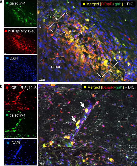
Dual-fluorescence co-immunostaining analysis of DEspR and Galectin-1 expression. a Analysis of the expanding tumor zone of a U87-CSC xenograft tumor invading through the tumor fibrous cap. Co-localization (yellow, yellow dotted circle) of increased human-DEspR expression (red dotted circle) in invasive U87 tumor cells and Galectin-1 (green dotted circle) is observed in the invasive front. {}, invasive tumor front. Host subcutaneous tissue is to the upper right corner. b Co-localization of hDEspR and Galectin-1 is detected in tumor cells adhering to the outer wall of a microvessel in the subcutaneous tissue demonstrating invasive nature of U87 in xenograft tumors and homing to microvessels
