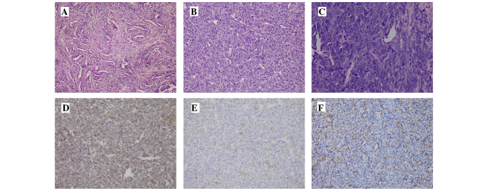
Figure 1.
Pathology and immunohistological staining of the removed tumor and metastatic lesions. (A) The tumor at first surgery appeared biphasic, containing spindle cells and numerous glandular structures lined by well-differentiated cuboidal epithelium (H&E; magnification, ×100). (B and C) The recurred tumor in the right neck at the second and third surgeries had a poorly differentiated appearance, characterized by predominantly undifferentiated round cell morphology [(B) H&E, magnification, ×100; (C) H&E, magnification, ×200]. (D) The tumor cells were diffusely positive for B-cell lymphoma-2. (E) The glandular structures showed weak positivity for cytokeratin. (F) The glandular structures were positive for vimentin. H&E, hematoxylin and eosin.

