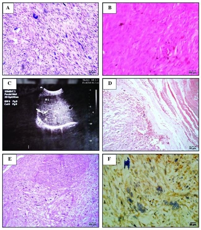
Figure 1.
Assessment of the biopsy samples at various stages. (A) The first prostate biopsy slice, stained with H&E, revealed tumor cells were predominantly spindle-shaped, the cytoplasm was dyed in red, nuclei were rod-shaped and rounded at each end. The majority of the tumor cells were with mild to moderate atypia and occasionally giant tumor cells were visible (magnification, ×20). (B) A biopsy slice of the first recurrence tumor, stained with H&E, revealed that tumor cells were in long spindle-shaped or oval and arranged in a palisade. Significant cellular atypia was observed and the cytoplasm was dyed in bright red. Mitotic cells were easily detected and a section of the tumor cell exhibited large nuclei, and interstitial was more loose (magnification, ×20). (C) The right bib metastatic cancer observed by the abdominal B-ultrasound results (a solid mass ~2.2×1.7 cm) in the capsule between diaphragm and liver. (D) A biopsy slice of the first resection of the rib metastases, stained with Masson-Goldnen trichrome, revealed red tumor cells were red (magnification, ×20). (E) A biopsy slice of the first resection of the rib metastases, stained with H&E (magnification, ×10). (F) A biopsy slice of the second resection of rib metastases, stained with actin antibody (magnification, ×40).

