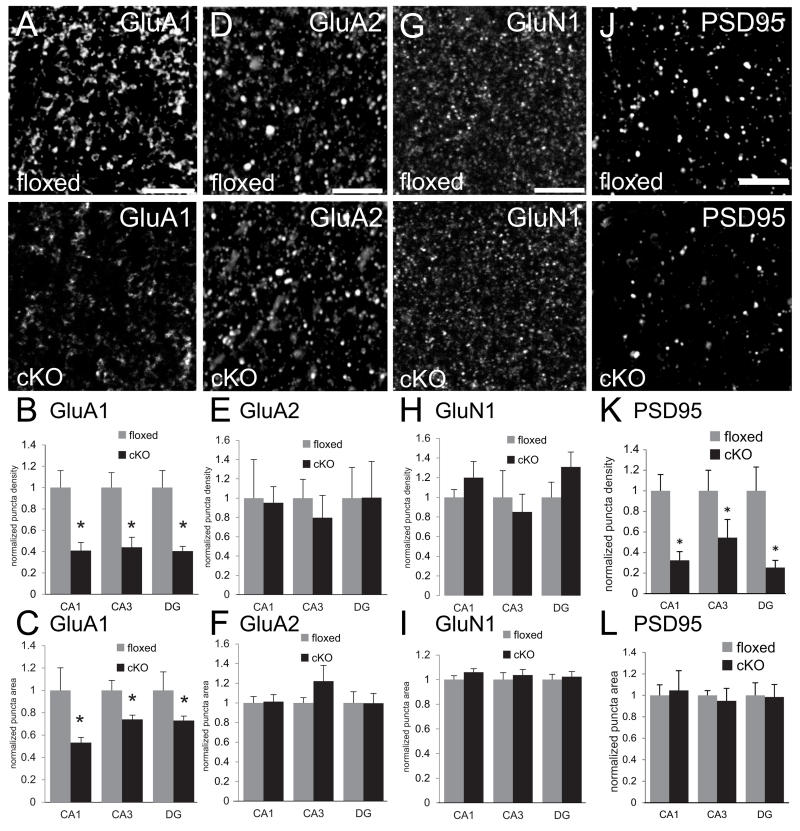
Fig. 5. Diminished density of GluA1 and PSD95 immunolabeled puncta in adult hippocampus of N-cadherin cKO mice.
(A,D,G,J) Representative confocal microscope images of sections through stratum radiatum of CA1 of adult floxed control mice (top row) or cKO mice (bottom row) immunofluorescently labeled for AMPAR subunits GluA1 (A), GluA2 (D), NMDAR subunit GluN1 (G), or the scaffolding protein PSD95 (J). Bars= 5 μm.
(B,E,H,K) Quantitative analysis of density of immunofluorescently labeled puncta. In all hippocampal subfields, the density of puncta immunolabeled for GluA1 (B) and PSD95 (K) was significantly lower in adult cKO mice (black bars) in comparison with that in adult floxed control mice (grey bars). In contrast, there were no differences between genotypes in density of GluA2 (E) or GluN1 (H). *p < 0.05, unpaired Student’s t-test; n=8 per genotype, both sexes. For each region, values were normalized to those of the floxed control mice for that region.
(C,F,I,L) Quantitative analysis of sizes of immunofluorescently labeled puncta. GluA1 immunolabeled puncta were significantly smaller in all hippocampal subfields in cKO mice (C, black bars) in comparison with floxed control mice (C, grey bars). There were no differences in sizes of puncta immunolabeled for GluA2 (F), GluN1 (I) or PSD95 (L). *p < 0.05, unpaired Student’s t-test; n=8 per genotype, both sexes. For each region, values were normalized to those of the floxed control mice for that region.

