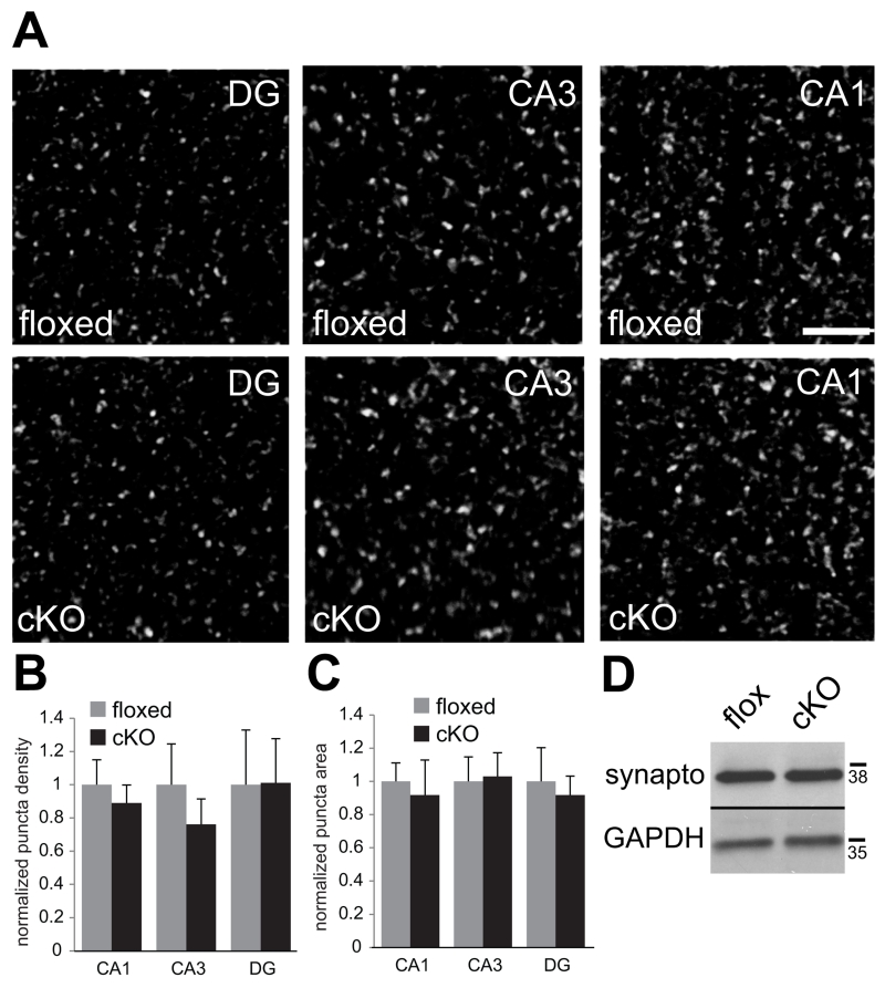
Fig. 7. No changes in levels or localization of presynaptic molecules in N-cadherin cKO mice.
(A) Representative confocal images showing vGlut1/2 immunolabeling in the indicated subfields of hippocampus taken from adult floxed control mice (top row) or cKO mice (bottom row). Bar= 5 μm.
(B and C) Quantitative analysis of vGlut1/2 puncta density (B) and size (C). n=8 mice per genotype, both sexes (p > 0.1, unpaired Student’s t-test). For each region, values were normalized to those of the floxed control mice for that region.
(D) Representative immunoblot of whole-hippocampal lysate showing similar levels of synaptophysin in floxed and cKO mice. GAPDH was used as a loading control. Numbers (on right) indicate approximate positions of molecular mass markers (kDa).

