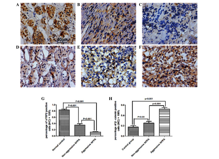
Figure 3.
Protein expression was assessed by IHC. sFRP2+ cells were detected in (A) normal pituitary tissues, (B) non-aggressive NFPAs and (C) aggressive NFPAs. β-catenin+ cells were detected in (D) normal pituitary tissues, (E) non-aggressive NFPAs and (F) aggressive NFPAs. Images G and H represent the quantitative analysis of IHC for sFRP2+ and β-catenin+ cells, respectively. Scale bar, 40 µm. Data are presented as the mean ± standard error of the mean. sFRP2, secreted frizzled-related protein 2; IHC, immunohistochemistry; NFPA, nonfunctioning pituitary adenoma.

