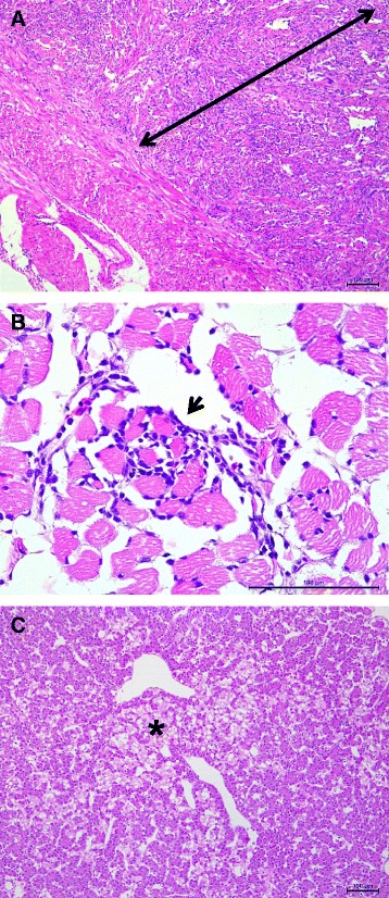
Fig. 3.

Photomicrographs showing the main microscopic findings in farmed Chilean coho salmon Oncorhynchus kisutch with HSMI-like disease. Panel a: Heart section stained with haematoxylin and eosin (H&E) showing infiltration of the spongious layer (arrow) by mononuclear cells (scale bar = 100 μm). Panel b: Red muscle section stained with H&E showing a focal infiltration of mononuclear cells among muscle fiber bundles (arrow head) (scale bar = 100 μm). Panel c Liver section showing focal necrosis of hepatocytes (asterisk) close to a blood vessel (scale bar = 100 μm)
