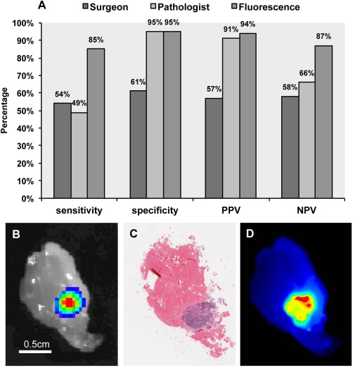
Figure 1.

Fluorescence localization of disease is compared to histopathological assessment and surgeon assessment using luciferase imaging as gold standard. (A) Sensitivity, specificity, positive predictive value (PPV), and negative predictive value (NPV) are shown for surgeon, pathologist, and fluorescence assessment of tumour wound‐bed margins compared to bioluminescence, which served as the gold standard. Representative images are shown of the margins assessed for (B) bioluminescence, (C) haematoxylin and eosin staining, and (D) matching fluorescence acquisition.
