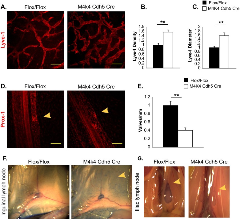
FIG 2.
Lymphatic abnormalities in Map4k4 Cdh5 Cre mice. (A to C) Ear skin from p6 Flox/Flox or Map4k4 Cdh5 Cre animals was stained with Lyve-1 as a marker of lymphatic capillaries. (A) Representative images. Scale bars, 100 μm. (B) Vessel density as a measure of percent stained area normalized to the Flox/Flox area. (C) Average vessel diameter as normalized to the Flox/Flox diameter. (D and E) Mesenteries from p2 Flox/Flox or Map4k4 Cdh5 Cre animals were stained with Prox-1 as a marker of lymphatic valves. (D) Representative images. The arrowheads indicate valves. Scale bars, 100 μm. (E) Numbers of valves per millimeter mesentery as normalized to Flox/Flox animals. (F and G) p16 pups were injected with Evans blue dye in the footpad, and the mice were sacrificed to visualize dye in lymph nodes 1 h later. The images are representative of at least 4 animals per genotype. (F) Inguinal lymph nodes; the arrowhead indicates enhanced capillary visualization in the skin of Map4k4 Cdh5 Cre mice. (G) Iliac lymph nodes; the arrowheads indicate a lack of dye in iliac lymph nodes of Map4k4 Cdh5 Cre mice. The error bars represent standard errors of the mean. **, P < 0.005; n = 4 to 6.

