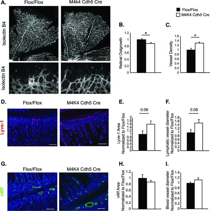
FIG 3.
Vascular abnormalities in Map4k4 Cdh5 Cre mice. (A to C) Retinas were isolated from p6 Flox/Flox or Map4k4 Cdh5 Cre pups and stained with isolectin B4. (A) Representative images (×5 magnification, scale bars = 250 μm [top]; ×20 magnification, scale bars = 50 μm [bottom]). (B) Quantitation of retinal outgrowth as the diameter from the optic nerve to the perimeter as normalized to Flox/Flox (*, P < 0.05; n = 5 or 6). (C) Vessel density quantified as percent stained area and normalized to Flox/Flox (*, P < 0.05; n = 6 or 7). (D to I) Intestines were isolated from p18 Flox/Flox or Map4k4 Cdh5 Cre pups. (D to F) Intestinal cross sections were stained with Lyve-1 as a lymphatic capillary marker and with 4′,6-diamidino-2-phenylindole (DAPI) to visualize villi. (D) Representative images; scale bars, 50 μm. (E) Quantitation of Lyve-1-stained area as normalized to Flox/Flox animals. (F) Quantitation of Lyve-1-stained-vessel diameter as normalized to Flox/Flox animals (n = 4 to 6). (G to I) Intestinal cross sections were stained with vWF as a blood endothelial marker and with DAPI to visualize villi; scale bars, 50 μm. (G) Representative images. (H) Quantitation of vWF-stained area as normalized to Flox/Flox animals. (I) Quantitation of vWF-stained-vessel diameter as normalized to Flox/Flox animals (n = 4 to 6). The error bars represent standard errors of the mean.

