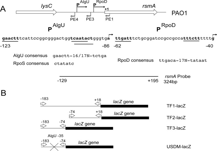
FIG 1.
Depiction of the rsmA genomic region, the promoters controlling rsmA expression, and the reagents used in this study. (A) Schematic representation of the rsmA genomic region. The sequences below the schematic represent those of the two potential promoters. The bent arrows above the sequence indicate the transcriptional start sites identified by primer extension analysis. The primers used in the primer extension experiments are identified by bent arrows below the genomic schematic. The potential promoters are indicated by a line underneath the sequence and are in bold. Potential sigma factor consensus sequences are indicated below the sequence. The probe used for RNase protection assays is indicated below, and position numbers are relative to the rsmA translational start site. (B) The rsmA-lacZ transcriptional fusions constructed are indicated. Arrows indicate the primers used in the construction of the transcriptional fusions. The position numbers above the arrows are in relation to the rsmA translational start codon. The X indicates the site-directed mutagenesis of the putative AlgU −35 consensus sequence.

