
FIG 3.
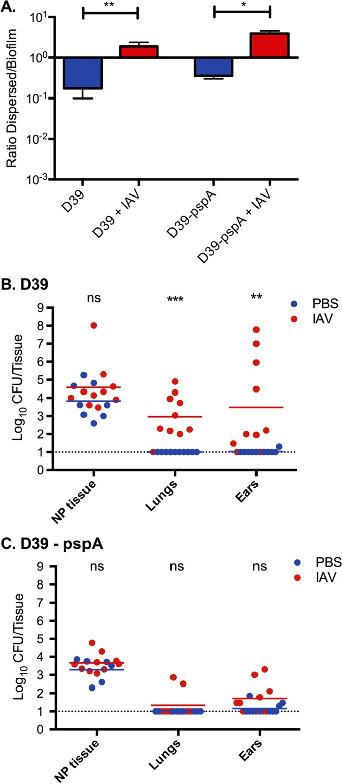
Role of PspA in biofilm release and survival in tissue after IAV infection. (A) D39 and D39 pspA null bacteria biofilms were made on top of respiratory epithelial cells for 48 h, and the cells were infected with IAV for 24 h. The ratio of dispersed (released) biofilm bacteria in the medium to the remaining bacteria in the biofilm was measured in the presence or absence of IAV. The bars show ratios with standard deviations. (B and C) Mice were colonized for 48 h with D39 wild-type bacteria and D39 lacking PspA and infected with 40 PFU of IAV. After 24 h the bacterial burden was measured in nasopharyngeal (NP) tissue and lung tissue and in the middle ears. D39 bacteria caused pneumonia and otitis media after IAV infection, whereas D39 pspA null pneumococci, even though they were released equally well in vitro (panel A), survived poorly in the lungs and middle ear tissues. Each experiment was performed twice with 5 mice in each group. Each dot in the graphs represents a single mouse. The lines represent the geometric mean. Statistics for all data sets were done using a Mann Whitney test (ns, not significant; *, P < 0.05; **, P < 0.01; ***, P < 0.001).