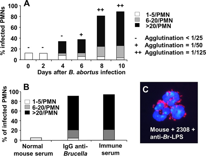
FIG 3.

Mouse PMNs phagocytize B. abortus after antibodies are generated. CD-1 mice were infected intraperitoneally with 106 CFU; then, mouse blood was collected at different days of infection and incubated with B. abortus 2308-RFP (MOI of 50) for 1 h. Blood smears were then fixed and mounted with ProLong Gold Antifade reagent with DAPI. At least 50 PMNs were counted per sample, and the number of intracellular bacterial in each PMN was determined. The relative agglutination titer for the day evaluated after intraperitoneal injection with B. abortus is indicated at the top of the bars according to the legend on the figure. (B) Mouse PMNs of CD-1 mice were incubated with B. abortus 2308-RFP at an MOI of 50 for 2 h and under different conditions of opsonization. Then, the number of phagocytized bacteria was recorded by fluorescence microscopy. (C) CD-1 mouse PMNs incubated with B. abortus 2308-RFP and anti-Brucella mouse serum at an MOI of 100. The image was cut from the microscope field, contrasted, and saturated using hue tool to obtain suitable color separation (magnification, ×200). Similar results were obtained with C57BL/6 and BALB/c mice.
