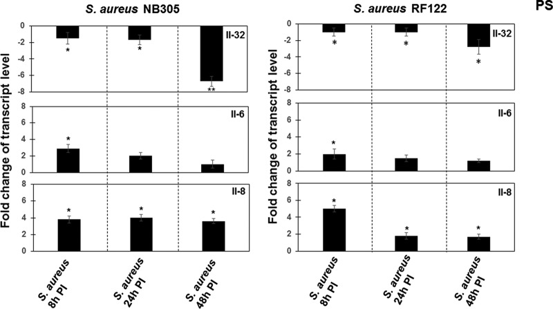
FIG 2.
Analysis of IL-32, IL-6, and IL-8 expression in S. aureus-infected cells. PS cells (2 × 10⁵) were grown for 24 h. The cells were then exposed to S. aureus NB305 or RF122 strains (MOI, 80:1) for 8 h, 24 h, and 48 h. Isolation of total RNA, synthesis of cDNA, and qRT-PCR were performed as described in Materials and Methods. After normalization using PPIA and RPL19 genes, interleukin expression at 8 h, 24 h, and 48 h p.i. was calculated relative to the values obtained from mock cells, arbitrarily set to 1. When the expression was decreased compared to that in unstimulated mock cells, data were presented as negative values. Data were calculated from three different experiments performed in triplicate. P values of <0.05 (*) and <0.01 (**) were considered significant.

