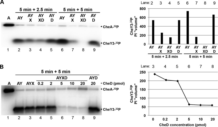
FIG 2.
CheD enhances phosphatase activity of CheX. Shown are phosphorylation-dephosphorylation assays using purified recombinant CheA (A), CheY3 (Y), CheX (X), and CheD (D) proteins. (A) Twenty picomoles of CheA was autophosphorylated using 10 μCi of [γ-32P]ATP (lane 1). CheA-32P was then incubated separately with 160 pmol of CheY3 for 5 min (lanes 2 and 6). Radiolabeled CheY3-32P was incubated with 0.15 pmol of CheX for 2.5 min or 5 min (lanes 3 and 7, respectively). The CheY3-32P and CheX reaction mixture was incubated with (lanes 4 land 8) or without (lanes 5 and 9) 15 pmol of CheD. The arrowheads indicate the positions of CheA-32P and CheY3-32P. (B) CheX phosphatase activity was efficiently enhanced by CheD. CheA was autophosphorylated as described above (lane 1). CheA-32P was then incubated with 160 pmol of CheY3 for 10 min (lane 2). The radiolabeled CheY3-32P was mixed with 0.15 pmol of CheX for 5 min without CheD (lane 3) or with 0.2 pmol (lane 4), 2 pmol (lane 5), 5 pmol (lane 6), 10 pmol (lane 7), or 20 pmol (lane 8) of CheD. CheY3-32P was also incubated with 20 pmol of CheD to confirm that CheD did not interfere with CheY3-P dephosphorylation (lane 9). The arrowheads indicate the positions of CheA-32P, CheY3-32P, and CheD. The relative intensities (“PI volumes”) of CheY3-32P are shown on the right with the corresponding phosphorimage lane numbers above.

