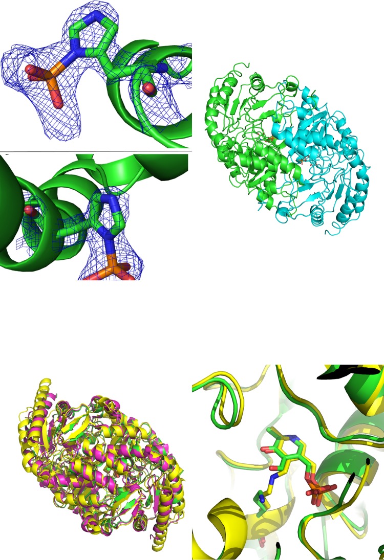
FIG 2.
(Upper left) 2mFo-DFc electron density maps for the phosphohistidine groups observed in the X-ray diffraction data for KES23458 at 1.5 σ. Diagrams show His31 (top) and His360 (bottom). (Upper right) Dimeric form of KES23458. One monomeric subunit is shown in green and the other in cyan, with PLP shown in each active site. (Lower left) Overlay for KES23458 from Pseudomonas sp. strain AAC, shown in green. The protein 4A6T from Chromobacterium violaceum is shown in yellow. The protein 4B98 from Pseudomonas aeruginosa is shown in magenta. (Lower right) The two proteins, KES23458 and 4A6T, overlaid, with the free PLP and Lys288 from KES23458; the internal aldimine from 4A6T is represented by sticks.

