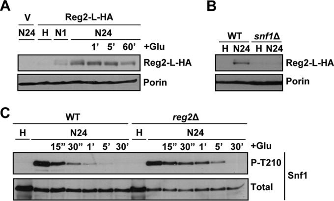
FIG 8.

Reg2 accumulation increases after prolonged glucose deprivation in a Snf1-dependent manner. (A) reg2Δ cells transformed with the vector pRS316 (V) or pReg2-L-HA (Reg2-L-HA) were grown to mid-log phase in selective SC medium lacking uracil and containing 2% glucose (H, high glucose) and then shifted to an otherwise identical medium containing no glucose for either 1 h (N1) or 24 h (N24). After 24 h of glucose starvation, the cultures were replenished with 2% glucose (+Glu) for either 1 min (1′), 5 min (5′), or 60 min (60′). (B) Cells of the indicated genotypes carrying pReg2-L-HA (Reg2-L-HA) were grown to mid-log phase in selective SC medium lacking uracil and containing 2% glucose (H, high glucose) and then shifted to an otherwise identical medium containing no glucose for 24 h (N24). In panels A and B, the levels of tagged Reg2 expressed from the native REG2 promoter (Reg2-L-HA) and Por1 protein (Porin) were detected by immunoblotting as for Fig. 7E. (C) Cells of the indicated genotypes were grown to mid-log phase in YEP medium containing 2% glucose (H, high glucose) and shifted to an otherwise identical medium containing no glucose for 24 h (N24). Following 24 h in no glucose, the cells were replenished with 2% glucose (+Glu) for either 15 s (15″), 30 s (30″), 1 min (1′), 5 min (5′), or 30 min (30′). The levels of phospho-Thr210-Snf1 (P-T210) and total Snf1 protein (Total) were analyzed by immunoblotting. The strains were MMY9 (reg2Δ), MKY323 (WT), and MKY366 (snf1Δ).
