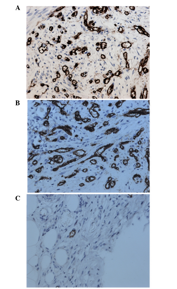
Figure 3.

Representative images of immunohistochemical analysis of CD34 in endometriotic lesions of the (A) control, (B) saline and (C) rapamycin-treated groups. Staining was performed with the streptomycin avidin-peroxidase method. Magnification, ×400.
