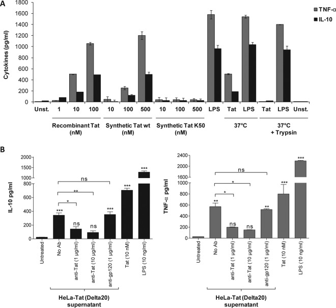
FIG 1.
The Tat protein induces TNF-α and IL-10 production in human monocytes in a specific manner. (A) Human monocytes were incubated with increasing amounts of full-length HIV-1 Tat protein, either recombinant (Tat), chemically synthesized (synthetic Tat), or acetylated at lysine 50 (K50). As a control, the recombinant Tat protein (10 nM) or LPS (100 ng/ml) was pretreated with trypsin for 1 h at 37°C or kept at 37°C for 1 h without trypsin. After 24 h of treatment, levels of TNF-α (gray bars) and IL-10 (black bars) in the supernatants of the cells were quantified by ELISA. Unst., unstimulated cells. (B) Human monocytes were incubated with HeLa-Tat cells (Delta20 line) stably transfected with tat-rev-vpu gene-conditioned medium (100 μl) in the absence or presence of a mixture of three monoclonal antibodies (1 μg or 10 μg/ml), recognizing epitopes localized in domains 1-15, 46-60, and 78-86 of the Tat sequence, or 10 μg/ml of a monoclonal antibody against HIV-1 gp120 (Mab110-4), which recognizes an epitope within the V3 region. As positive controls, human monocytes were treated with either recombinant Tat (10 nM) or LPS (10 ng/ml). After 24 h, the cell supernatants were collected, and TNF-α (gray bars) and IL-10 (black bars) levels were quantified by ELISA. The data represent means and SD for three independent experiments. Statistical significance was analyzed by one-way ANOVA follow by Bonferroni posttests and is marked as follows: *, P < 0.05; **, P < 0.01; ***, P < 0.001; ns, nonsignificant. All bars were compared to those for untreated cells unless otherwise specified (indicated with a black line above the bars).

