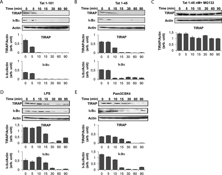
FIG 4.
HIV-1 Tat protein leads to time-dependent and proteasome-dependent TIRAP/MAL degradation via its N-terminal domain. THP-1 cells (106 cells/condition) were treated with recombinant GST-Tat (full-length; Tat 1-101) (A) or the GST-tagged N-terminal fragment (Tat 1-45) (B) at 180 nM. (C) Cells were preincubated with MG132 (20 μM) for 60 min before treatment with Tat 1-45. As positive controls, cells were treated with LPS (1 μg/ml) (D) and Pam3CSK4 (1 μg/ml) (E). Cells were lysed after different times (0 to 90 min). Cellular proteins were separated by SDS-PAGE and analyzed by Western blotting for TIRAP/MAL and IκBα expression. Actin was also quantified as a loading control. Quantification of the bands obtained from 3 independent experiments was performed using ImageJ software. Data represent TIRAP/MAL expression or IκB expression normalized to actin expression.

