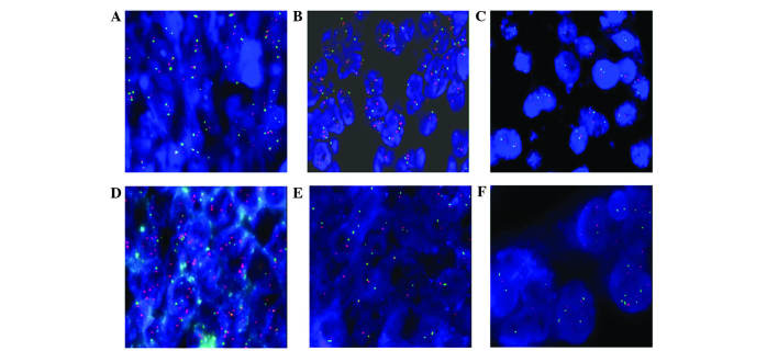
Figure 1.
SPOP gene alterations and numerical abnormalities of chromosome 17 in ovarian tissue. (A) In normal ovarian tissue, signals/nucleus of CEP17 all showed two green spots, and signals of SPOP gene showed two red spots. (B) In certain ovarian cancer tissues, signals/nucleus of CEP17 showed two green spots, and signals of SPOP gene showed two red spots. (C) Deletion of the SPOP gene had a SPOP/CEP17 signal ratio of <0.7. (D) Amplification of the SPOP gene had a SPOP/CEP17 signal ratio of >2.0. (E) Monosomy of CEP17 had a CEP17 signals/nucleus ration of <1.5 in >10% cells. (F) Polysomy of CEP17 had a CEP17 signals/nucleus ration of >2.5 in >10% cells. SPOP, speckle-type POZ (pox virus and zinc finger protein) protein; CEP17, chromosome enumeration probe 17.

