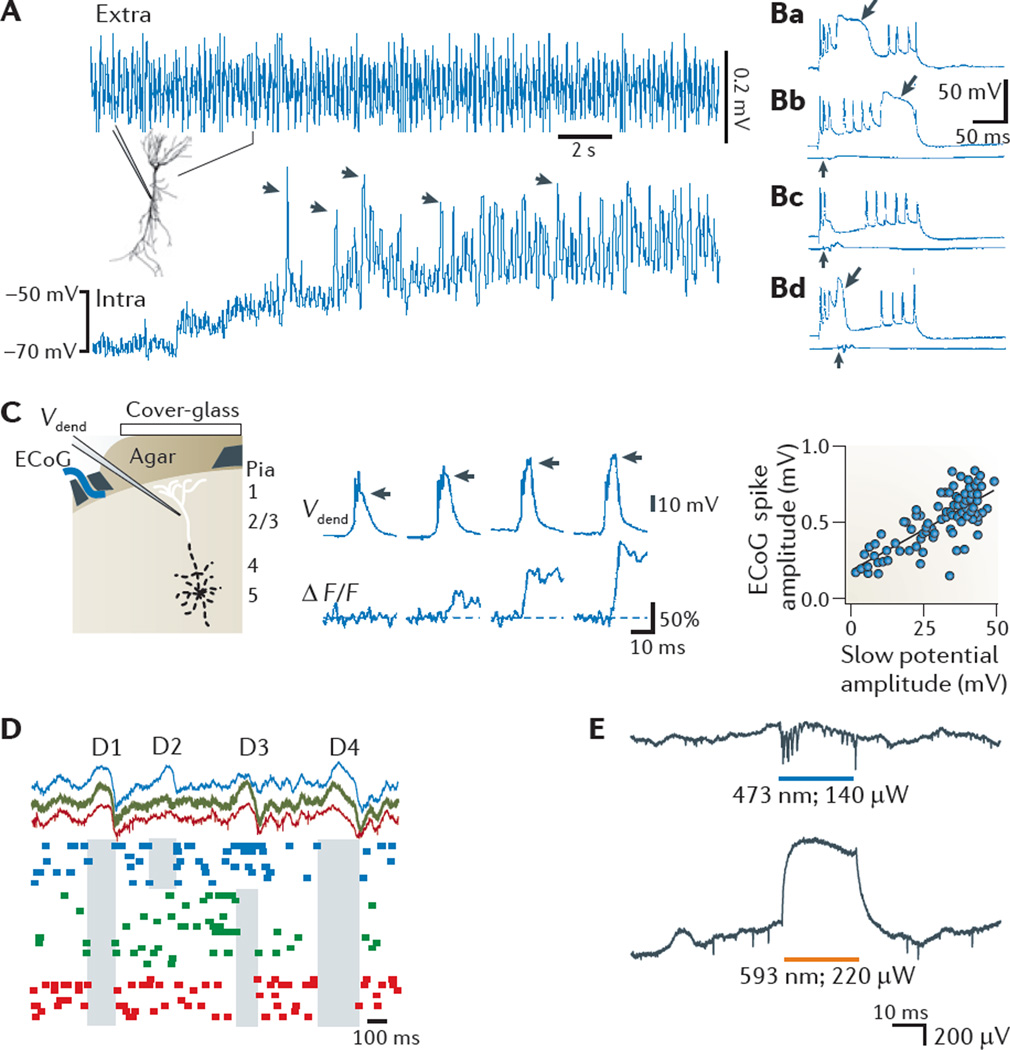
Figure 3. Non-synaptic contributions to the LFP.
Ca2+ spikes, disfacilitation and disinhibition contribute to the local field potential (LFP). A | Voltage-dependence of a theta-frequency oscillation in a hippocampal pyramidal cell dendrite in vivo. A continuous recording of extracellular (extra) and intradendritic (intra) activity in a hippocampal CA1 pyramidal cell is shown. The holding potential was manually shifted to progressively more depolarized levels by intradendritic current injection. The recording electrode contained QX-314 to block Na+ spikes. Note the large increase in the amplitude of the intradendritic theta oscillation upon depolarization. Arrows, putative high-threshold Ca2+ spikes phase-locked to the LFP theta oscillation. Ba | Dendritic Ca2+ spikes (shown by an arrow) have a large amplitude and are long-lasting in vivo. Bb–Bd | The response of a CA1 pyramidal cell to ventral hippocampal commissural stimulation (vertical arrows) paired with dendritic depolarization. Such inhibition can delay (Bb), prevent (Bc) or abort (Bd) the dendritic Ca2+ spike. LFPs recorded from a nearby electrode in the pyramidal layer show the timing and magnitude of the stimulation (lower traces in Bb–Bd). Note that the number of Na2+ spikes remains approximately the same, irrespective of the presence or absence of the Ca2+ spike. C | Whisker stimulation-evoked dendritic Ca2+ spikes correlate with surface cortical LFP changes. The setup for recording the electrocorticogram (ECoG), intradendritic potential (Vdend) and Ca2+ fluorescence is shown in the left panel. The relationship between the intradendritic potential amplitude (horizontal arrows) and simultaneously measured Ca2+ influx (ΔF/F) is shown in the middle panel. The ECoG response as a function of the Ca2+ spike (‘slow potential’) amplitude is shown in the right panel. D | ‘Down’ states in cortical pyramidal cells during sleep produce extracellular LFP ‘delta’ waves. Shown are simultaneously recorded LFP (top) and unit activity (bottom) at three layer 5 intracortical locations (spaced approximately 1 mm apart; indicated by different colours). Note that down states (shaded areas), reflected as positive waves (delta waves) in the LFP, can be either strongly localized (in D2 and D3) or more widespread (in D1 and D4). E | Generation of extracellular potentials by depolarization or hyperpolarization of a limited number of CA1 neurons that express both channelrhodopsin 2 (ChR2) and halorhodopsin, in response to blue (top) and yellow (bottom) light in vivo. Note the depolarization-induced negative LFP (top) and the hyperpolarization-induced positive LFP (bottom) in the pyramidal layer. Part A is reproduced, with permission, from REF. 159 © (1998) Wiley. Part B is reproduced, with permission, from REF. 160 © (1996) National Academy of Sciences. Part C is reproduced from REF. 161 © (1999) Macmillan Publishers Ltd. All rights reserved. Part D is reproduced, with permission, from REF. 56 © (2005) Cambridge Journals. Part E courtesy of E. Stark, New York University, Langone Medical Center, USA.

