
Figure 4.
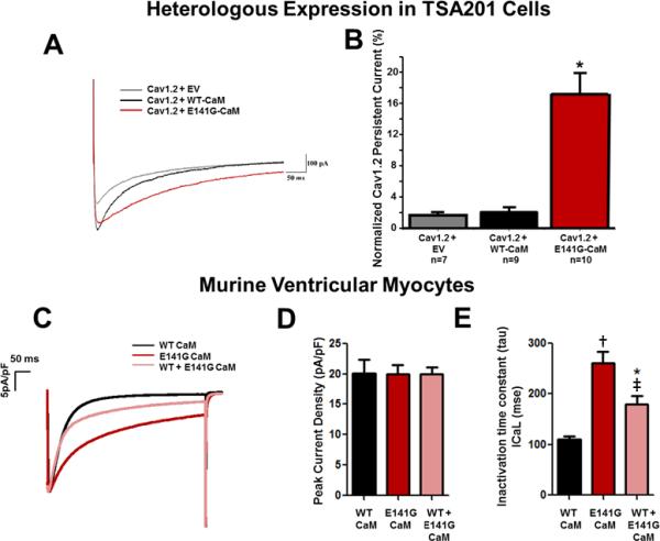
E141G-CaM leads to increased CaV1.2 persistent current and alters current inactivation without affecting peak current density in murine ventricular myocytes. (A) Representative tracings of persistent CaV1.2 current from CaV1.2+EV, CaV1.2+WT-CaM and CaV1.2+E141G-CaM determined from a holding potential of −90 mV to +30 mV with 500ms duration in TSA201 cells. (B) Group data showing CaV1.2 late current normalized to peak (%) for CaV1.2+EV, CaV1.2+WT-CaM, and CaV1.2+E141G-CaM in TSA201 cells. *P<0.05 vs. CaV1.2+WT-CaM. (C) Representative examples of traces for each experimental group in murine ventricular myocytes. (D) Average current densities (pA/pF) obtained in cells dialyzed with WT- or E141G-CaM in murine ventricular myocytes. (E) Effect of E141G-CaM alone or mixed with 75% WT CaM on inactivation time constant of the CaV1.2 compared to WT-CaM in murine ventricular myocytes. Data are mean ± SD. (n=7 *P<0.05; †P<0.001 vs. WT-CaM. ‡P<0.01 vs. E141G-CaM alone).