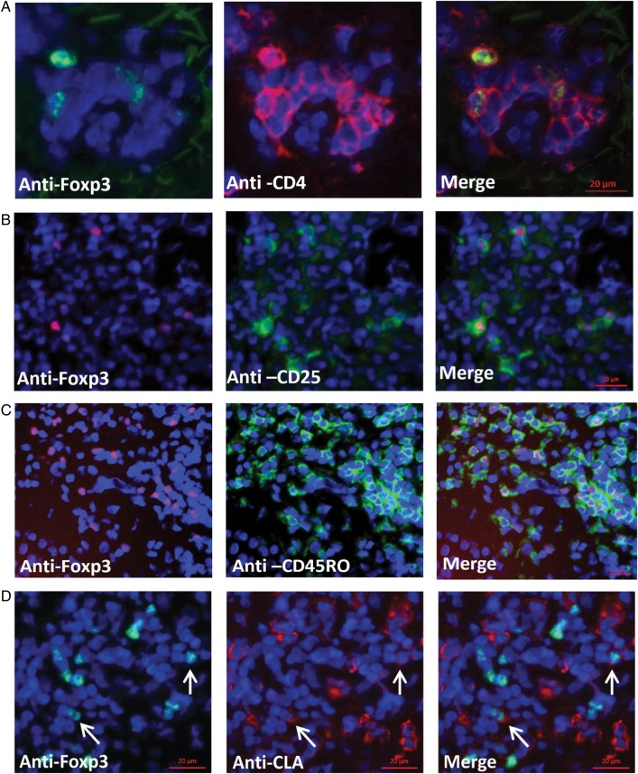
Figure 1.
Characterization of Foxp3+ cells during human herpes simplex virus type 2 reactivation. A–D, Frozen genital skin biopsy specimens from an acute genital lesion were stained for Foxp3 (green) and CD4 (red; A); Foxp3 (red) and CD25 (green; B); Foxp3 (red) and CD45RO (green; C); and Foxp3 (green) and cutaneous lymphocyte antigen (CLA) (red; D). Arrows point to Foxp3+CLA+ cells. All sections were also counterstained with DAPI (blue). Scale bar = 20 µm.

