Fig. 1.
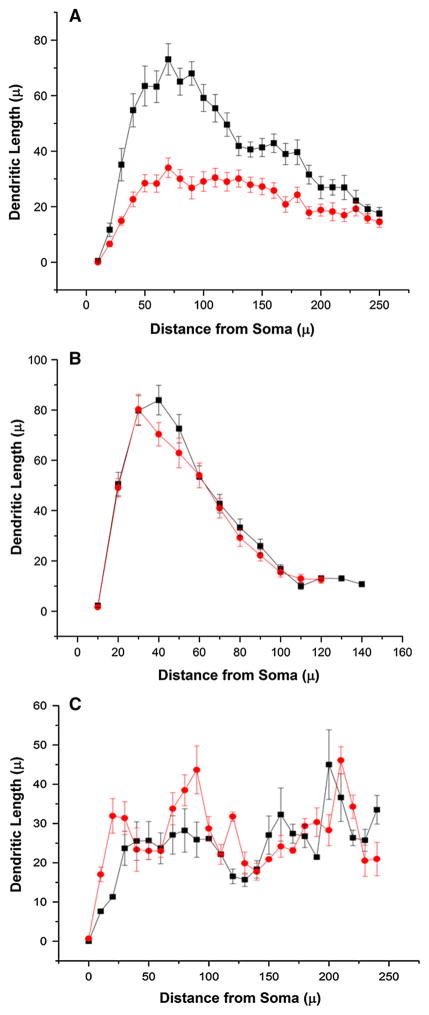
Sholl analysis of apical and basal dendrites in 2 and 8 weeks post-CLP-exposed mice and shams. a Sholl analysis of apical CA1 dendrites of the hippocampus demonstrates significant loss of arbors in animals 8 weeks post-CLP or sham (five mice in each group, neurons = 29 in each group; D = 0.5, P <0.01, Kolmogorov–Smirnov test). b Sholl analysis of basal CA1 dendrites demonstrates comparable arbors in animals 8 weeks post-CLP or sham (four mice in each group, neurons = 27 in sham and 26 in CLP; D = 0.1, NS, Kolmogorov–Smirnov test). c Sholl analysis of apical CA1 dendrites of the hippocampus demonstrate comparable arbors in animals 2 weeks post-CLP or sham (six mice in each group, neurons = 6; D = 0.3, NS, Kolmogorov–Smirnov test). Sham black, CLP red (Color figure online)

