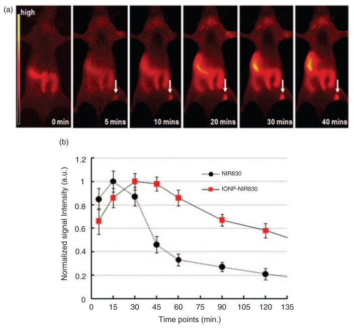
Figure 5.
Near infrared (NIR) images at different time points before and after administered IONP-NIR830 (a) and signal intensity profiles in sentinel lymph nodes (SLNs) tracked by either free NIR830 or IONP-NIR830 (b). Data at each time point were averaged from those collected from six animals. The signal intensities arbitrary unit (a.u.) were normalized based on the highest signals for both groups. The absolute signal intensity of free NIR dye (3800 a.u.) was approximately twice higher than that of IONP-NIR830 (2010 a.u.). Reduction of the NIR signal intensity in IONP-NIR830 is due to the quench effect from the aggregation of NIR830 dyes on the iron oxide nanoparticles (IONP) surface. It is noticed that increased signal intensity in the right abdominal area is likely due to the accumulation of IONP-NIR830 nanoparticles in the liver after cleared out of the lymph nodes via lymphatic drainage.

