Figure 1.
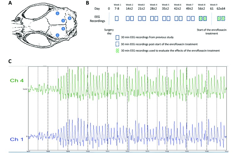
(A) Schematic diagram illustrating the positioning of the epidural recording screw electrodes (1 through 4) and reference electrode (R). (B) Experimental design. EEG recording (30-min session twice weekly) began on day 7 or 8 after surgery and continued for the following 7 wk. On day 61, the drinking water was replaced with enrofloxacin-containing water (1.7 mL of enrofloxacin 25 mg/mL in 750 mL of drinking water), and additional EEG recordings were performed on days 62 and 64. The enrofloxacin treatment was stopped on day 65. (C) The clonic seizure of rat 5 started on day 64 after 308 s of EEG recording and lasted for 40 s. The intensity of the clonic seizure was classified as stage 4 (clonic seizure in a seating position).11 The EEG recordings presented SWD that were similar in amplitude (0.3 to 0.7 mV) and frequency (average, 7 SWD per s) to that observed during absence seizure.

