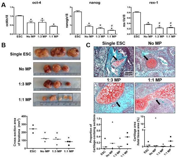
Fig. 6.
Effect of mineral particles on the pluripotency of ESC aggregates. (A) Real-time PCR analysis of pluripotent gene expression. *P<0.05 vs. single ESCs (n=4). (B) Equal cell numbers of single ESCs (ESC suspension harvested directly from 2D culture), no MP, 1:3 MP, or 1:1 MP ESC aggregates were subcutaneously injected in NOD SCID mice and the tissue masses were harvested 28-days post-injection and examined macroscopically. Black rectangle indicates the three individual pieces of teratoma derived from a single injection of 1:1 MP aggregates. The cross-section area of teratomas were quantified by Image J. *P<0.05 vs. single ESCs. (C) Teratoma sections were stained by safranin O/fast green in order to visualize the positive-stained cartilage tissues (red) within each section. Representative images of stained teratomas sections showed the presence of cartilage tissues as indicated by arrows (Scale bar = 100 μm). The proportion of cartilage-containing sections and the cartilage cross-sectional area % were calculated based on histomorphometric analyses.

