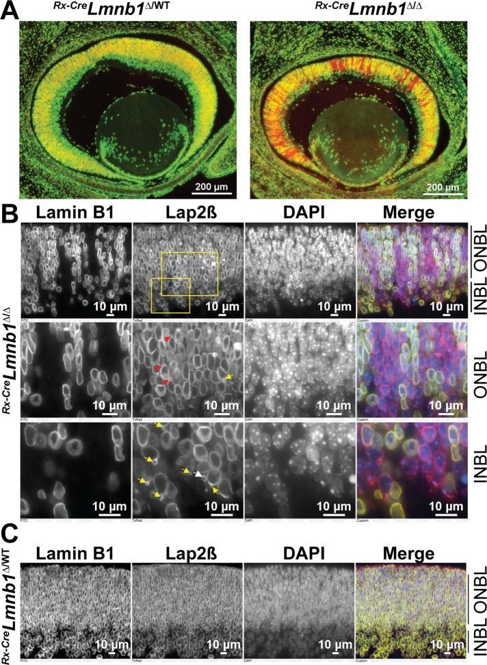
FIGURE 1:
Lamin B1 deficiency in mouse embryonic retina results in the formation of nuclear blebs. (A) Lamin B1 immunofluorescence pattern (green) in E14.5 Rx-CreLmnb1Δ/WT (left) and Rx-CreLmnb1Δ/Δ (right) embryonic retinas counterstained with DAPI (pseudocolored in red). Owing to the variegated expression of the Rx-Cre transgene, there is a mosaic pattern of Lmnb1 inactivation in the retina, resulting in columns of lamin B1–expressing and lamin B1–deficient cells. (B) Immunostaining of E14.5 Rx-CreLmnb1Δ/Δ retinas with antibodies for lamin B1 and Lap2β. In the merged image, lamin B1 is green and Lap2β is red; DNA was stained with DAPI (blue). Top, overview; middle, zoomed-in view of ONBL; bottom, zoomed-in view of inner INBL. Red and yellow arrows point to apical and basal nuclear blebs, respectively. White arrow points to a solitary bleb within the INBL. See also Supplemental Figure S1. (C) Identical immunohistochemistry studies on retinas from E14.5 Rx-CreLmnb1Δ/WT littermate control mice.

